
How is thyroid and parathyroid ultrasonography performed and when is your doctor order a thyroid ultrasonography ?
In Thyroid and parathyroid ultrasound sound waves are being used to produce a series of thyroid gland images, whereby a radiologist will determine the shape and size of the gland in the body; Hypo- and hyperthyroidism and related conditions may alter thyroid gland size and shape with consequent problems.Therefore, when the doctor diagnoses the complications of thyroid disease, he/she will request the thyroid ultrasonography. This imaging modality will be selected when the doctor wants to evaluate your thyroid tissue and check for thyroid nodules. Ultrasonography provides real-time images and instant information such that the physician can use this technique for other necessary medical procedures, including thyroid biopsy. Thyroid ultrasonography does not require any special preparation and you do not need to do anything special before doing it, just put on a comfortable clothing and go to the ultrasonography office or center.
What is thyroid ultrasonography?
Thyroid ultrasonography is safe and painless procedure, The modality creates images of internal body organs using sound waves and the procedure is performed by moving a transducer (prob) directly on ultrasound gel impregnated skin. During ultrasound imaging the sound waves or ultrasound pulses are transmitted directly to the body by prop and through gel, and the reflections or echo of these pulses off the body organs are recorded and displayed as an image.It does not use ionizing radiation (X-rays), so the patient is not exposed to radiation, and since the imaging is performed in real-time mode, it can show blood flow in vessels and depicts movements and structure of internal organs.
How is thyroid ultrasonography performed?
In Thyroid ultrasonography procedure, the patient rests comfortably on the bed, and the physician moves a special, gel-coated ultrasound probe, on the surface of the skin. As the radiologist performs the ultrasonography, the echo of sound pulses is recorded in the device and the physician can observe the ultrasound image of the target on a display monitor. This procedure does not cause any pain or harm to the patient, and also it does not lead to complications of radiation, so pregnant patients can use this imaging modality confidently and safely.
Thyroid and its function in the body
The ultrasonography examination in this area creates an image of the thyroid gland and the surrounding tissues. This butterfly-shaped gland is one of nine endocrine glands in the body which is located in front of the neck and above the clavicle bone. It is composed of two lobes connected by a bridge.
You can see the shape of this gland in the picture.
This gland produces and releases hormones into the bloodstream
The hormone secreted by this organ called the thyroid hormone and regulates the function of different organs of the body. One of the most common thyroid conditions which occur in 5 to 10% of the general population is thyroid nodules or masses. Sometimes these nodules are palpable through skin but in some cases they are not palpable and radiologists diagnose them by other examinations and tests.
Thyroid ultrasonography is a highly sensitive examination, and most impalpable nodules are detected in ultrasound. It can be diagnosed in 70% of adults’ ultrasonography. The vast majority of thyroid nodules are benign and pose no risk to the individuals, but in some cases they are true thyroid tumors and require recognition and treatment.
When is Thyroid ultrasonography performed?
The most common uses and indications of thyroid ultrasound:
- To detect mass(es) in the thyroid or its surrounding structures. Sometimes patient or physician notice a protrusion in the neck or find symptoms suggesting a thyroid nodule.In this condition, thyroid ultrasound can detect probable thyroid nodule in the gland, confidently.
- To assess the appearance of thyroid nodules and to evaluate their nature (benign or malignant) and whether a biopsy is needed or not. In case of biopsy it can be performed by Fine Needle Aspiration (FNA) under ultrasound guide which increases the biopsy accuracy.
Examination of thyroid masses and nodule structures can help the physician in identification of their nature as benign or malignant and also their grades. If thyroid ultrasound results suspect to a malignant mass, the doctor will use a thyroid biopsy in the next step. In this procedure a fine needle is directed to target area under ultrasound guidance and sampling is performed precisely in desired location by local anesthesia.
Other indications
- To detect and evaluate possible nodules impalpable in physical examination. The presence of more nodules in the thyroid may suggest multinodular goiter in the patient. So the radiologist identifies the possible malignant nodules by physical examination and ultrasonography to find more information about their multiplicity.
- To evaluate the increase in nodule numbers that have changed significantly over a given period of time.
Because of real-time feature in this technique, it can be used in guide-required procedures including FNA and Core Needle biopsy. It also can be used along with other devices and instrumentation procedures (diagnostic and therapeutic evacuation procedures).
Ultrasound pulses are being produced and detected simultaneously during the procedure, so it has the capability for guidance of biopsy needle to a target location, for example a thyroid nodule. This feature of ultrasound is also being used in minimally invasive procedures including microwave and radiofrequency ablation of thyroid nodules . In these cases, the radiologist can treat thyroid masses without damaging the surrounding normal thyroid tissue. More information regarding these techniques is provided in other sections of the website.
Preparation for thyroid and parathyroid ultrasound (Thyroid ultrasonography)
Ultrasonography doesn’t need any special preparation and a comfortable clothes is all you need to perform it.Children who are preparing for thyroid ultrasound typically are agitated and stressful, so explaining the steps to them and bringing a toy, storybook or something that can be used as distraction is helpful.
Since ultrasonography produces no pain, you can discuss it to your child and assure them about its safety. During the procedure entertain and distract with your children.
How does it work?
In fact, in thyroid ultrasound and any other kind of ultrasonography sound waves and calculations of their reflection or echo time is being used to produce an image. This is similar to what bats do to detect obstacles in their path; they calculate the distance to an object by sending ultrasound waves and calculating the return or reflection time.These waves can be used to calculate the shape, distance and the nature of objects (filled with liquid or not).These reflections and echoes produce real-life images of organs and limbs; the physician can identify abnormalities by evaluation of images.
What are the Benefits and Risks of Ultrasound?
Benefits:
- Most ultrasound examinations are non-invasive procedures (without needles or injections)
- Ultrasonography is a painless procedure, it is easy to perform, accessible and cheaper than other modalities.
- This modality is very safe and does not use any ionizing radiation (X-rays).
- It produces a clear image of soft tissue incomparable with other techniques such as radiography.
- Ultrasound provides a real-time image which is a very useful feature guiding invasive procedures such as needle biopsy and fluid aspirations.
Risks:
- There is no known or reported deleterious effect of ultrasonography for patients.
Equipped with the most up-to-date technological advances and innovative procedures and with the collaboration of experienced radiologists and interventionists leader in their specialties, Tirad Ultrasound and Imaging Center is providing cutting edge, innovative and minimally invasive/non-invasive procedures for diagnosis and treatment of most benign and malignant thyroid gland conditions. We continuously working to provide patients with the world’s most recent advances in diagnostic imaging and minimally invasive treatments.
Post

RFA Thyroid – The newest cure for thyroid cancer
No Comments
What is Thyroid Radiofrequency Ablation – How are RF used in the treatment of thyroid cancer? Thyroid Radiofrequency Ablation (RFA) is a minimally invasive method for destruction of benign thyroid…
Read More
What is a Thyroid Biopsy? How is a thyroid biopsy performed?
What is a Thyroid Biopsy? How is a thyroid biopsy performed? You may have thyroid problems or have people around you with this conditions. In some cases, your doctor or…
Read MoreRF Consultation (Radiofrequency)
RF Consultation (Radiofrequency) We talk about RF consultation . In the Radiofrequency Applications for Thyroid ( Thyroid RF ) Diseases section, general points regarding radiofrequency technology along with specific details…
Read More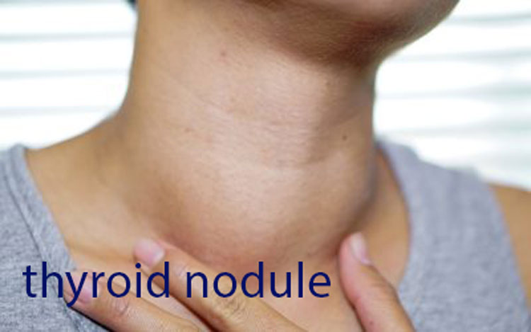
What Is A Thyroid Nodule?
What Is A Thyroid Nodule? Are All Thyroid Nodules Serious And Must Be Treated? Have you ever heard of thyroid nodules? Do you know the thyroid gland? Thyroid is a…
Read More